Chest imaging
ABCDEF: Airways/quality, Bones, Cardiac silhouette, Diaphragm, Effusions, Fields/foreign bodies.
How to read a film
Read every chest radiograph the same way, every time. The ABCDEF pass is slower than “glance and guess,” and it catches the missed pneumothorax, the malpositioned tube, and the small effusion hiding on the lateral.
- A
Assess quality, airways, always compare
Rotation: spinous processes bisect the clavicular heads. Inspiration: 9–10 posterior ribs. Penetration: vertebral bodies visible through the heart. Airways: narrowing, deviation, foreign body. Always pull the prior film.
- B
Bones and soft tissue
Subcutaneous air, rib fractures, cervical ribs. Soft-tissue swelling can be the only clue to a missed trauma film.
- C
Cardiac silhouette, mediastinum, hilum
AP films enlarge the heart. Cardiothoracic ratio >50% suggests cardiomegaly on a PA film only. Widened mediastinum is generally >8 cm — first exclude rotation.
- D
Diaphragm and gastric bubble
Left hemidiaphragm is usually lower. Flattening is sensitive for emphysema. A missing gastric bubble on the left is a hint, not a diagnosis.
- E
Effusions
Blunting of the costophrenic angle. Small fluid is seen first on the lateral. Upright PA misses fluid until >250 mL; recumbent films are worse. Lateral decubitus: free-flowing fluid layers, loculated fluid does not.
- F
Fields and foreign bodies (lines)
Focal vs diffuse: PTX, pneumonia, edema. Silhouette sign = a density against a normal border. ETT tip 3–5 cm above the carina.
↑ intrapleural pressure pushes the trachea (PTX). ↓ intrapleural pressure pulls it (atelectasis / mucus plug).


Trachea, push vs pull

Pushes (↑ intrapleural pressure)
- Tension pneumothorax
- Large pleural effusion
- Massive hemothorax
- Large mass
Pulls (↓ volume)
- Lobar atelectasis / mucus plug
- Fibrosis / pneumonectomy
- Scarring after TB
- Unilateral hypoplasia
Pattern recognition on CXR


| Sign | Means | Where to look |
|---|---|---|
| Silhouette sign | Pulmonary density touching a normal border erases it | RML erases the right heart; RLL erases the diaphragm; lingula erases the left heart |
| Air bronchogram | Patent airway inside consolidated lung | Pneumonia > atelectasis; also pulmonary edema, hemorrhage |
| Kerley B lines | Thick interlobular septa | CHF, lymphangitic spread |
| Deep sulcus | Supine PTX collecting anterior-caudal | ICU films — do not wait for an upright |
| Bat-wing / perihilar | Central alveolar filling | Cardiogenic edema, DAH, PCP (bat-wing GGO on CT) |
CT chest — windows and patterns
CT is for an abnormal CXR you cannot characterize, or a normal CXR you do not believe. HRCT (<2 mm, non-contrast) is the study for diffuse parenchymal disease: inspiratory, expiratory, supine, and prone.
- Scout first — anatomy and tubes at a glance.
- Soft-tissue window: mediastinum, heart, vessels. LV > RV normally. Main PA diameter should be less than the ascending aorta; PA ≥29 mm has PPV 97% for pulmonary hypertension.
- Lung window: parenchyma. Inspiratory HRCT: round trachea. Expiratory HRCT: D-shaped trachea; air trapping declares itself.
- Coronary calcium is visible even on non-contrast studies — mention it.
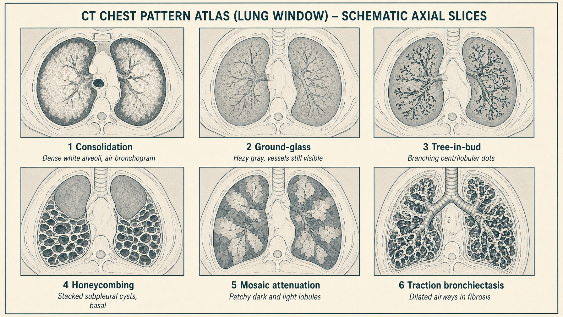
| Pattern | What you are seeing | First etiologies |
|---|---|---|
| Consolidation | Complete alveolar filling; dense attenuation that obscures vessels ± air bronchograms | Pneumonia > malignancy |
| Ground-glass (GGO) | Partial filling; hazy ↑ attenuation that does not hide architecture | Infection, edema, blood, fibrosis, malignancy |
| Tree-in-bud | Filled centrilobular bronchioles — branching dots | Infection including TB, CTD, ABPA, CF, tumor emboli |
| Emphysema / bleb | Airspace >1–2 cm; bleb is the smaller cousin | COPD, congenital, spontaneous PTX |
| Mosaic attenuation | Patchy ↑ and ↓ attenuation from air trapping, poor perfusion, or GGO. Expiratory images worsen air trapping | Hypersensitivity pneumonitis, obstruction, CTEPH, infection, edema |
| Honeycombing | Stacked cystic airspaces. Microcystic <4 mm, macrocystic >4 mm | NSIP often <4 mm; IPF / UIP often >4 mm |
| Subpleural reticulation | Septal lines ~1 cm from the pleura | UIP > NSIP; can be aging |
| Bronchiectasis | Irreversibly dilated airways; tram-tracks, signet-ring | Post-infectious, CF, NTM, traction from fibrosis |
| Atelectasis vs effusion | Atelectasis enhances with contrast and can improve prone; effusion is homogeneous | Do not call both without a decubitus or CT |